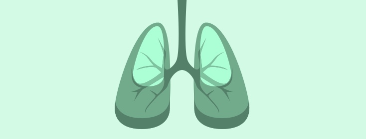
Respiratory 101: The Respiratory Tract
It's helpful for anyone with a respiratory disease like COPD to at least have a basic understanding of the respiratory system and how it works. Today we're going to take you on a fun and educational ride through the respiratory tract. We promise to keep this an easy read for your educational enjoyment.
Ready? Let's go!
The air we breathe contains about 21% oxygen. If we were to put eyes on one of these oxygen molecules and follow it (at slow motion speed) we'd learn quite a bit about the anatomy of the respiratory tract. (Grab fake eyes. Place on oxygen molecule).
Air (along with our oxygen molecule with fake eyes) first enters the nose. It passes through hairs, although some pathogens and particles in the air are not so lucky. The nose is the first passage of the upper respiratory tract. It acts to filter air, and to warm and humidify it.
Turbinates
If you could feel what our oxygen molecule was feeling, you'd notice the air around us was nice and toasty. As our molecule uses it's fake eyes to peer from one side to the other, you'll see three boney, shelf-like projections on either side. These are the superior, middle, and inferior conchae. Together they are called turbinates, and they act as humidifiers and heaters to humidify and warm inhaled air to body temperature, which so happens to be a toasty 98.7° Fahrenheit or 37° Celsius.
Mucus membrane
This is what we will see covering all cells lining the respiratory tract. It's a sticky substance meant to trap any particles that make it into airways. It also helps to keep cells lining airways from getting dehydrated.
Cilia
These are small, hairlike structures that move back and forth. They line the respiratory tract from the nose all the way to the respiratory bronchioles. They work as an escalator to move mucusdown your upper airway to the back of your throat where they can be swallowed. Stomach acids will then destroy the germs. In the lower airways they move mucus up to the back of the throat to be swallowed. (Sure, if you like, you can also spit them out. Either method is acceptable).
Goblet cells
These are cells randomly scattered along the walls of the respiratory tract that secrete mucus. Particles and pathogens are balled up in mucus and moved to the back of the throat by cilia. When you have lung diseases like asthma or COPD, you may have an elevated number of these cells, meaning you produce excessive mucus.
Pharynx
It begins at the back of the nose where it is called the nasopharynx. It then takes a turn downward to the back of the mouth to the uvula. You are now in a region called the oropharynx. Here oxygen shares a passage with food. It travels beyond the base of the tongue and approaches a gate-like object called the...
Glottis
You watch as the glottis swings open, and the food travels down the esophagus. Sputum brought to the back of your throat by cilia also moves down the esophagus. As the glottis closes our oxygen molecule is allowed to pass by and into the lower airway.
Larynx
It is quite sturdy, as it is lined with nine cartilages. If you take your hand and feel the front of your neck, you can feel these cartilages. What you are touching is the upper portion of your larynx, and it is sometimes called the Adam's Apple. As our oxygen continues on (and looking through our fake oxygen eyes) we can see a white band on either side. These are the vocal cords. As our person inhales, the cords are open and allow air (and our oxygen molecule) to easily pass through them.
Trachea
This is another large opening that is kept open by 16-20 c-shaped cartilages. It is often referred to as the windpipe. It is the last part of our travel through the upper airways.
Carina
This is a fork in the road. Our oxygen molecules can turn left and go to the left lung, or turn right to the right lung. Our molecule takes a left turn.
Bronchi
The carina is the beginning of the bronchi, or larger bronchi. Like the larynx and trachea, they are kept open by cartilage. We are also now inside the lungs. As we go down, these branch off into smaller and smaller passages. Our oxygen molecule eventually makes it to the smaller bronchi. (If you listen closely, you can hear the distant beating of the heart).
Bronchioles
The bronchi branch off into even smaller passages that are no longer surrounded by cartilage. Instead, they have smooth muscles wrapped around them. During COPD flare-ups or asthma attacks, this muscle constricts and squeezes or narrows these passages. Now, however, they are open and we travel easily through them.
Terminal Bronchioles
These are the smallest airways that do not come into contact with alveoli.
Respiratory Bronchioles
There are the smallest airways and they lead to alveolar ducts.
Alveolar ducts
These are very small passages that are connected to 10-20 air sacs.
Alveoli
These are air sacs, or balloon-like structures where gas exchange occurs. They are connected to a supply of venous capillaries. Because there are a lot of oxygen molecules in an alveoli, and only a few (if any) in venous capillary blood, oxygen molecules easily cross the alveolar-capillary membrane into the blood stream.
Lung Parenchyma
This is a term referring to the terminal bronchioles, respiratory bronchioles, and alveoli. When your immune system is compromised, which sometimes happens when you have a disease like COPD, it is this part of the lungs that becomes infected and inflamed. This is called pneumonia.
Arteries
This is blood that contains freshly oxygenated blood from the lungs. Our oxygen molecule binds with a hemoglobin molecule, which is shaped like an inner tube or small donut. When this happens, the blood turns a bright red. This bright red, freshly oxygenated blood then travels to one of the various cells of the body.
Cellular metabolism
Our oxygen molecule is used to make energy for a cell. The byproduct of cellular metabolism is carbon dioxide.
Now let's put our fake eyes on a carbon dioxide molecule...
Venous blood
Once oxygen leaves a hemoglobin molecule to a cell, the blood turns a darker, purplish-blue color. As oxygen leaves the blood, our carbon dioxide molecule enters the blood and binds to hemoglobin. This de-oxygenated, darker blood travels back to the lungs. As we follow along (looking through our fake eyes) we take a rollercoaster ride through the various passages to a venous capillary in the lungs.
Since there are more carbon dioxide molecules in venous blood than in alveoli, our carbon dioxide molecule easily crosses the alveolar-capillary membrane into an alveoli.
Air exchange units
In this way, alveoli is where ventilation takes place. When you inhale, oxygen enters the alveoli and into the blood stream. When you exhale, carbon dioxide leaves the alveoli and into the atmosphere. You are now exhaled back into the atmosphere. This is the end of the ride.
I hope you enjoyed our journey (our class).
Please return your fake eyes to the fake eye receptacle to your right. Any wisdom retained is free of charge. Coming soon to our anatomy theme park is a journey through the cardiovascular system. Stay tuned!

Join the conversation